Written by Mrs. Patsi Elisavet, Radiologist, Scientific Manager of the Anassa Computed Tomography Laboratory.
MRI and CT are standard methods of 3D imaging of tissues, organs and body parts for the diagnosis of different diseases.
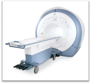
Magnetic tomography (MRI) is a diagnostic scanning technique based on the principles of magnetic resonance and uses a strong magnetic field of waves and radio frequencies without exposure to radiation. It enables the diagnostic approach of the Central Nervous System (brain, spine) with great accuracy in highlighting tumors, metastases, vascular malformations, neurodegenerative diseases.
It is an important weapon for neurosurgeons, orthopaedic surgeons and other interventional specialties for the strategy of surgery. It is the test of choice for the demonstration of ischemic stroke in the acute phase and for the diagnostic approach to joints, soft tissues of the body such as ligaments, menisci, tendons and bone marrow.
The specific techniques of MRI have an important role in urology, e.g. detection – diagnostic accuracy and staging of prostate cancer (multiparametric study) and possible early recurrence after treatment.
Similarly, specific MRI techniques have a similarly important role in the detection – evaluation of female pelvic and rectal cancer (staging – recurrence).
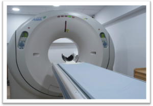
CT scan (CT) is a scanning technique based on the absorption of X-rays by body tissues and rendering sharp 3D images of them. A CT scan can provide detailed information about parts of the body such as bones, lungs, soft tissues, heart and blood vessels. It is used to diagnose and monitor various cases, with the following main indications:
Very good imaging of bones, e.g. fractures, as well as the digestive system and lungs, calcifications (calcium accumulations in the walls of blood vessels), while its use in emergency situations is important for the detection of acute abdominal diseases, acute strokes or bleeding.
Computed tomography (CT) imaging provides very accurate images of the coronary arteries of the heart, showing not only the narrowing of an artery but also the entire atherosclerotic plaque before it becomes narrowed.
The choice of MRI over CT depends on the part of the body the clinician wants to examine and on his diagnostic approach in consideration of the other clinical laboratory testing. For example, the doctor suggests a CT scan to show a bone fracture, an acute brain hemorrhage, especially a post-traumatic one, especially considering that the development of technology allows today to reduce the radiation exposure levels in the CT scanner.
On the other hand, if the clinician wants to examine pathology of ligaments, tendon fibers, spinal cord, he will suggest MRI.
At ANASSA, with the opening of a state-of-the-art 1.5 Tesla MRI and 128-slice CT scanner with minimal radiation dose, we have completed the creation of a complete Diagnostic – Imaging Center located within the clinic, with 24-hour operation, 365 days a year, serving both external and internal patients directly and qualitatively.